Hoof anatomy collection
Showing 25–48 of 48 results
Extreme case of laminitis print
Anatomical photograph by Dr. Christoph von Horst. The foundered hoof specimen shows the destruction of the coffin bone in a hoof with severe chronic laminitis. Scientific photoprint, size appr. 20 x 30cm / 8 x 12in on glossy Fuji Crystal … Continued
incl. 19% VAT
plus Shipping Costs
Add to cartEquine foal hoof vascularisation
Anatomical photograph of a horse hoof sectional anatomy specimen by Dr. Christoph von Horst. The anatomical blood vessel corrosion cast shows finest details of the hoof and distal limb blood vessel supply. Scientific photoprint, size appr. 20 x 30cm / … Continued
incl. 19% VAT
plus Shipping Costs
Add to cartEquine hoof selective impregnation (SI) print
Anatomical photograph of an SI sheet plastination specimen by Dr. Christoph von Horst. Detailed view of the deep digital flexor tendon and other tendon structures in the horse hoof. Scientific photoprint, size appr. 20 x 30cm / 8 x 12in … Continued
incl. 19% VAT
plus Shipping Costs
Add to cartEquine distal limb anatomy print
incl. 19% VAT
plus Shipping Costs
Add to cartHorse hoof anatomy teaching chart
incl. 19% VAT
plus Shipping Costs
Add to cartHorse foot anatomy plastination print
incl. 19% VAT
plus Shipping Costs
Add to cartEquine distal limb anatomy print
incl. 19% VAT
plus Shipping Costs
Add to cartEquine laminitis coffin bone rotation
incl. 19% VAT
plus Shipping Costs
Add to cartEquine hoof anatomy plastination print
incl. 19% VAT
plus Shipping Costs
Add to cartLaminated hoof anatomy chart print
This “low-tech” heavy duty laminated image print is still an excellent choice for many occasions. Super solid and waterproof. You can draw and write on the lamination with a permanent marker and clean it with alcohol afterwards. Fits in any … Continued
incl. 19% VAT
plus Shipping Costs
Add to cartHorse navicular syndrome pathology
incl. 19% VAT
plus Shipping Costs
Add to cartHorse hoof anatomy plastination print
Anatomical photograph of a horse hoof sectional anatomy specimen by Dr. Christoph von Horst. The anatomical specimen shows finest details of the morphology. Print in size appr. 20 x 30 cm / 8 x 12 inches with printed signature as shown … Continued
incl. 19% VAT
plus Shipping Costs
Add to cartEquine laminitis pathology print
Image print showing characteristic signs of equine laminitis in a pathological wet specimen by Dr. Christoph von Horst. The colour preserved sectional anatomy specimen provides a natural view of a fresh cut hoof section showing the pathological patterns in the … Continued
incl. 19% VAT
plus Shipping Costs
Add to cartHorse Laminitis Foot CPE
incl. 19% VAT
plus Shipping Costs
Add to cartHorse hoof anatomy chart (german)
Anatomical photograph of a horse hoof cross section plastinate with labelled anatomical structures by Dr. Christoph von Horst. The anatomy specimen shows topography details relevant for equine podiatry and a better understanding of hoof diseases and hoofcare. Scientific photoprint, size appr. … Continued
incl. 19% VAT
plus Shipping Costs
Add to cartEquine hoof anatomy print
Anatomical photograph by Dr. Christoph von Horst. The anatomy specimen shows finest anatomical details. Scientific photoprint, size appr. 20 x 30cm / 8 x 12in on glossy Fuji Crystal Archive Paper (or similar high-quality photo paper). Watermark will not be shown … Continued
incl. 19% VAT
plus Shipping Costs
Add to cartLaminitis double hoof wall pathology
incl. 19% VAT
plus Shipping Costs
Add to cartEquine hoof vascularisation details
Anatomical photograph of a horse hoof sectional anatomy specimen by Dr. Christoph von Horst: Detail view showing the horse hoof vascularisation in a research outlet plastinate of a horse hoof. Blood supply of the coffin bone, navicular bone, suspensory apparatus … Continued
incl. 19% VAT
plus Shipping Costs
Add to cartNavicular bone Plastinate
Anatomical photograph by Dr. Christoph von Horst: Anatomy and vascularisation of the navicular region and the navicular bone in the horse. Blood vessels entering the navicular bone and the equine coffin bone which are relevant for typical problems like navicular … Continued
incl. 19% VAT
plus Shipping Costs
Add to cartEquine distal limb sectional anatomy hoof
Anatomical image print of an equine hoof anatomy specimen showing hoof care insights relevant not only for farriers by Dr. Christoph von Horst. Sectional anatomy of the horse foot with equine distal limb anatomy details like bones, tendons, etc. Scientific … Continued
incl. 19% VAT
plus Shipping Costs
Add to cartLaminitis anatomical hoof
Anatomical image print of an equine laminitis hoof anatomy specimen showing hoof pathology insights relevant not only for farriers by Dr. Christoph von Horst. Sectional anatomy of an equine foundered hoof shows changes in the anatomy of the coffin bone … Continued
incl. 19% VAT
plus Shipping Costs
Add to cartEquine laminitis coffin bone perforation print
Anatomical photograph of a horse hoof sectional anatomy specimen by Dr. Christoph von Horst. The anatomical specimen shows anatomical and pathological details of the horse hoof laminitis disease and the foundered hoof. The hoof sole was perforated by the coffin bone … Continued
incl. 19% VAT
plus Shipping Costs
Add to cartHorse canon bone section
Anatomical image print of a horse limb cross section showing equine anatomical insights relevant for everyone working with horses. Sectional anatomy of the cannon bone, musculus interosseum medius / suspensory ligament, deep flexor tendon, superficial flexor tendon, arteries, veins, nervs, … Continued
incl. 19% VAT
plus Shipping Costs
Add to cart
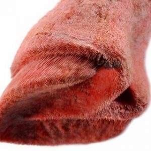
Horse foot blood vessels print
Anatomical photograph of a juvenile horse hoof blood vessel corrosion cast specimen by Dr. Christoph von Horst. The anatomical specimen shows finest details of the morphology and topography of the blood vessels in the equine hoof. The anatomical corrosion cast image … Continued
incl. 19% VAT
plus Shipping Costs
Add to cart