Deutsch
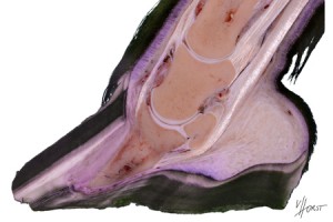 Equine Laminitis
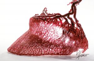
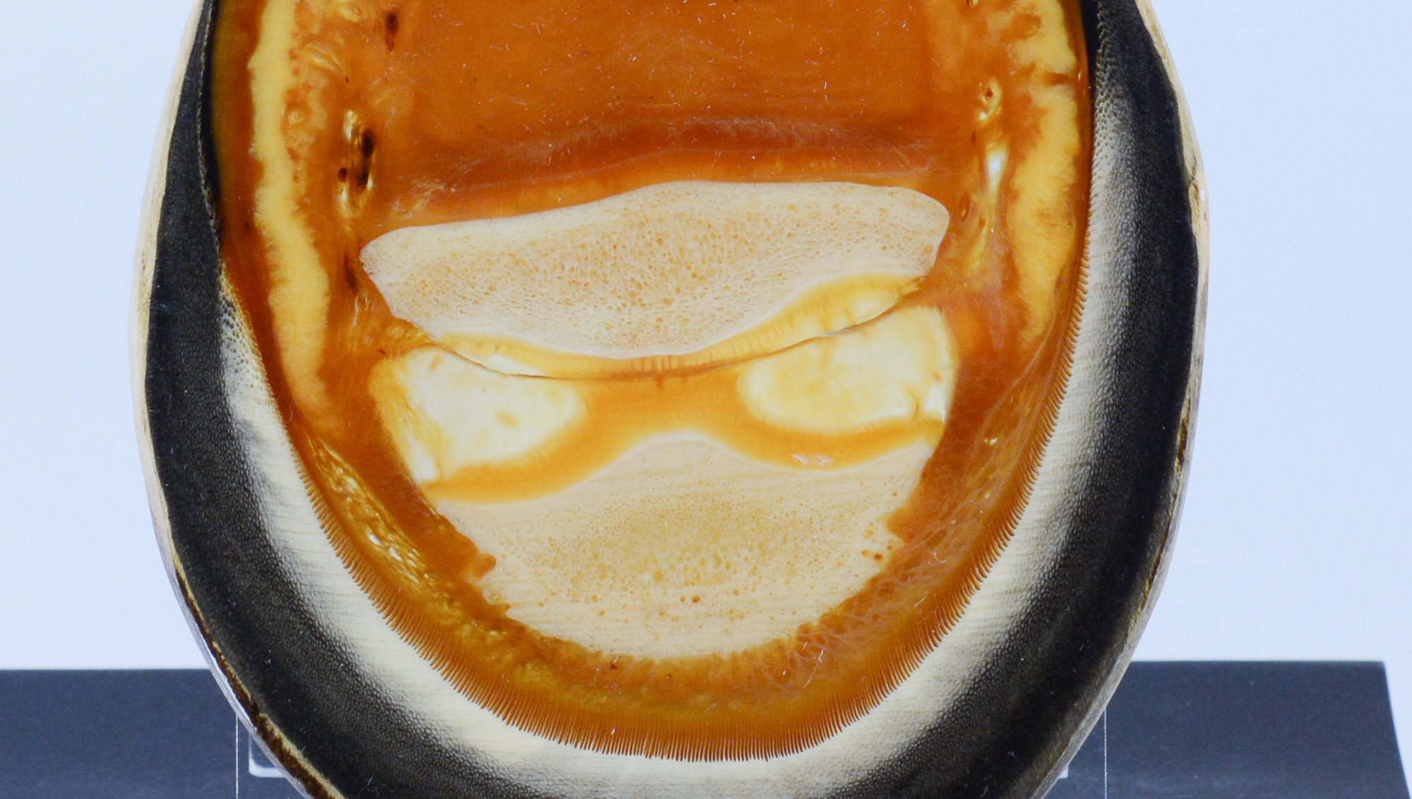

HC Biovision Dr. Christoph von Horst
Since the start of his dissertation in the year 2000, Dr. Christoph von Horst is working on plastination and anatomical preparation techniques. When he founded HC Biovision in 2004, high-end plastination services became available for museums, universities, teaching facilities and private individuals worldwide.