Deutsch
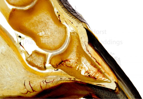
Description
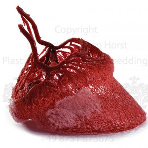
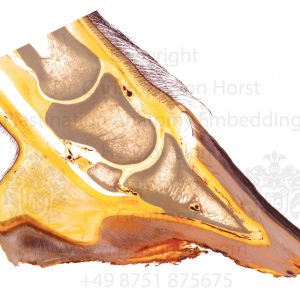
Anatomical photograph of a horse hoof sectional anatomy specimen by Dr. Christoph von Horst:
Detail view showing the horse hoof vascularisation in a research outlet plastinate of a horse hoof. Blood supply of the coffin bone, navicular bone, suspensory apparatus of the coffin bone, etc. The anatomical specimen shows finest details of the morphology.
Scientific photoprint, size appr. 20 x 30cm / 8 x 12in on glossy Fuji Crystal Archive Paper (or similar high-quality photo paper). Watermark will not be shown on the print. For other formats and materials including prints on canvas, alu dibond, acrylic, etc. please contact us directly.
Interested in real plastinates and anatomical specimens? Besides custom made plastinates and wet specimens we can also offer trial plastinates for budget prices. Please check our Offer Section.