Back to Image prints

Horse canon bone section

12,00 

5 in stock

Description

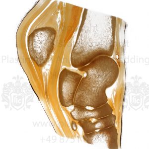
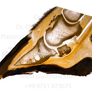
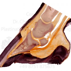
Anatomical image print of a horse limb cross section showing equine anatomical insights relevant for everyone working with horses. Sectional anatomy of the cannon bone, musculus interosseum medius / suspensory ligament, deep flexor tendon, superficial flexor tendon, arteries, veins, nervs, etc. Sheet plastination anatomical specimen if the equine distal limb by Dr. Christoph von Horst.

Scientific photoprint, size appr. 20 x 30cm / 8 x 12in on glossy Fuji Crystal Archive Paper (or similar high-quality photo paper). Watermark will not be shown on the print. For other formats and materials including prints on canvas, alu dibond, acrylic, etc. please contact us directly.

Interested in real plastinates and anatomical specimens? Besides custom made plastinates and wet specimens we can also offer trial plastinates for budget prices. Please check our Offer Section.

You may also like…

Equine foal hoof vascularisation

Anatomical photograph of a horse hoof sectional anatomy specimen by Dr. Christoph von Horst. The anatomical blood vessel corrosion cast shows finest details of the hoof and distal limb blood vessel supply. Scientific photoprint, size appr. 20 x 30cm / … Continued

12,00 

incl. 19% VAT

plus Shipping Costs

Add to cart
Equine hock joint anatomy print

Equine hock tarsal joint anatomy photograph by Dr. Christoph von Horst. T he veterinary anatomy specimen shows anatomical details of the horse hock joint and surrounding tendons, connective tissue, etc. Scientific photoprint, size appr. 20 x 30cm / 8 x 12in … Continued

12,00 

incl. 19% VAT

plus Shipping Costs

Add to cart
Equine head nasal sinus anatomy
[:de]Bestellungen im Online Shop sind bisher nur in englischer Sprache möglich.[:en]Anatomical picture print. Move mouse on the image below for maximum resolution view.[:]
12,00 

incl. 19% VAT

plus Shipping Costs

Add to cart